Show me a diagram of the human heart? Here are a bunch! Interactive Biology, with Leslie Samuel
In this interactive, you can label parts of the human heart. Drag and drop the text labels onto the boxes next to the diagram. Selecting or hovering over a box will highlight each area in the diagram. Download Exercise. Tweet. Rights: The University of Waikato Te Whare Wānanga o Waikato Published 16 June 2017 Referencing Hub media.

Heart Anatomy chambers, valves and vessels Anatomy & Physiology
Browse 8,896 human heart anatomy photos and images available, or search for human heart anatomy vector to find more great photos and pictures. Browse Getty Images' premium collection of high-quality, authentic Human Heart Anatomy stock photos, royalty-free images, and pictures.
Structure and Function of the Heart
A Labeled Diagram of the Human Heart You Really Need to See The heart, one of the most significant organs in the human body, is nothing but a muscular pump which pumps blood throughout the body. The human heart and its functions are truly fascinating. The heart, though small in size, performs highly significant functions that sustains human life.

Basic Anatomy of the Human Heart Cardiology Associates of Michigan Michigan's Best Heart Doctors
Images are labelled, providing an invaluable medical and anatomical tool. The test mode allows instant evaluation of user progress. Atlas of human anatomy: Left ventricle, Left atrium, Anterior papillary muscle Pericardium : Pericardial cavity, Transverse pericardial sinus, Oblique pericardial sinus
1 Labeled illustration of the human heart 1 [1]. This figure... Download Scientific Diagram
Anatomy of the human heart and coronaries: how to view anatomical structures. This tool provides access to an MDCT atlas in the 4 usual planes, allowing the user to interactively discover the heart anatomy. The images are labeled, providing an important medical and anatomical tool. The quiz mode makes it possible to evaluate the user's progress.
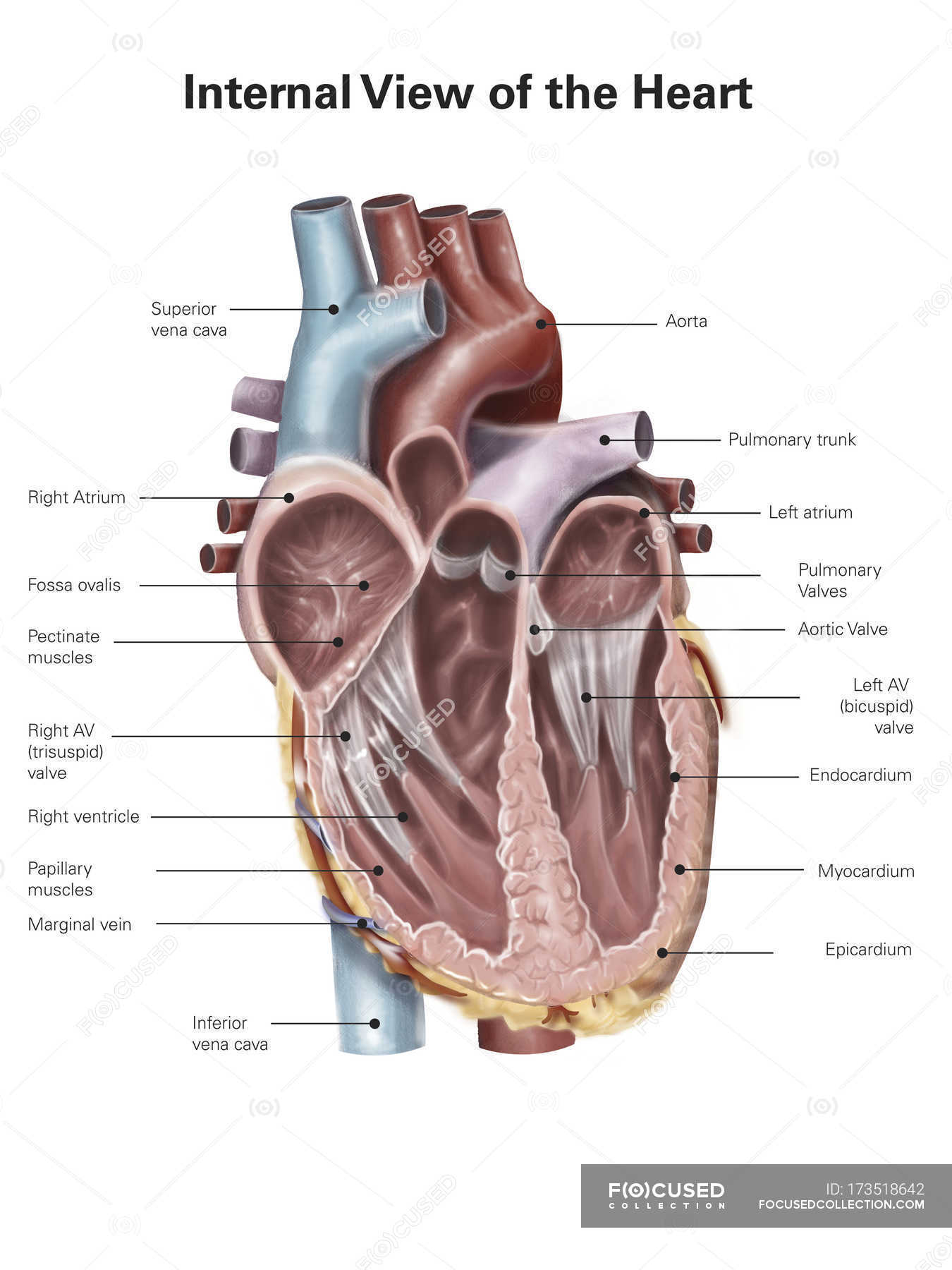
Human heart with labels — cross section, anatomy Stock Photo 173518642
Browse 700+ anatomy of the heart labelled stock photos and images available, or start a new search to explore more stock photos and images. Sort by: Most popular An hand drawn illustration of anatomy of the human heart with. Heart Blood Flow Circulation of blood through the heart. Cross Section of Heart with Labels on White Background

an image of the human heart with labels on it's main parts and functions
Browse 150 anatomy of the heart labelled photos and images available, or start a new search to explore more photos and images. Browse Getty Images' premium collection of high-quality, authentic Anatomy Of The Heart Labelled stock photos, royalty-free images, and pictures.
Human Heart Front View Interior Carlson Stock Art
152 Heart Anatomy With Labels Stock Photos, High-Res Pictures, and Images - Getty Images Browse Images Creative Images Browse millions of royalty-free images and photos, available in a variety of formats and styles, including exclusive visuals you won't find anywhere else. See all creative images Trending Image Searches Happy New Year
labeled diagram of heart
$9.99 Add To Cart Anatomy of the Heart Welcome to the anatomy of the heart made easy! We will use labeled diagrams and pictures to learn the main cardiac structures and related vascular system. In addition to reviewing the human heart anatomy, we will also discuss the function and order in which blood flows through the heart.
Heart And Labels Drawing at GetDrawings Free download
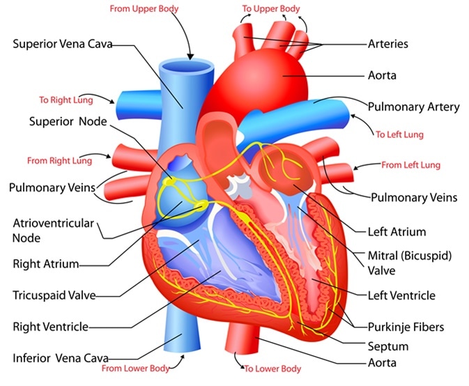
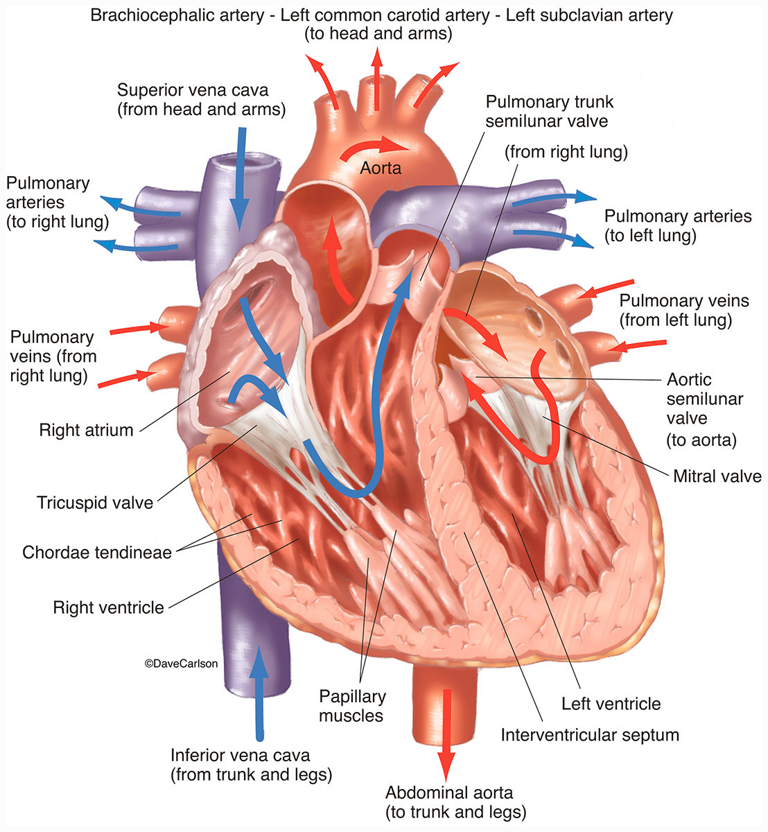
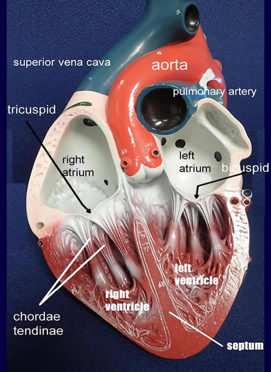
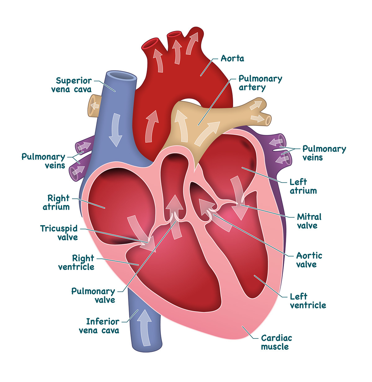
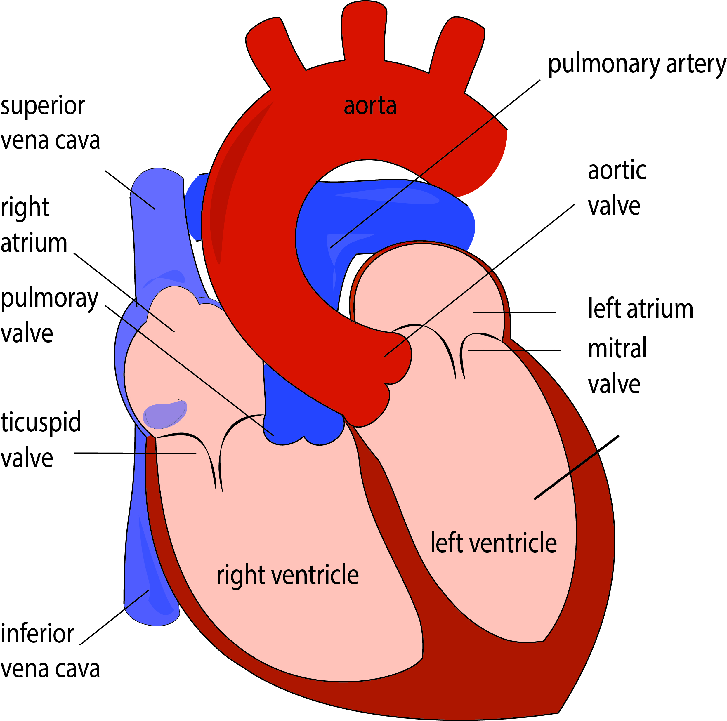
Well-Labelled Diagram of Heart The heart is made up of four chambers: The upper two chambers of the heart are called auricles. The lower two chambers of the heart are called ventricles. The heart wall is made up of three layers: The outer layer of the heart wall is called epicardium. The middle layer of the heart wall is called myocardium.
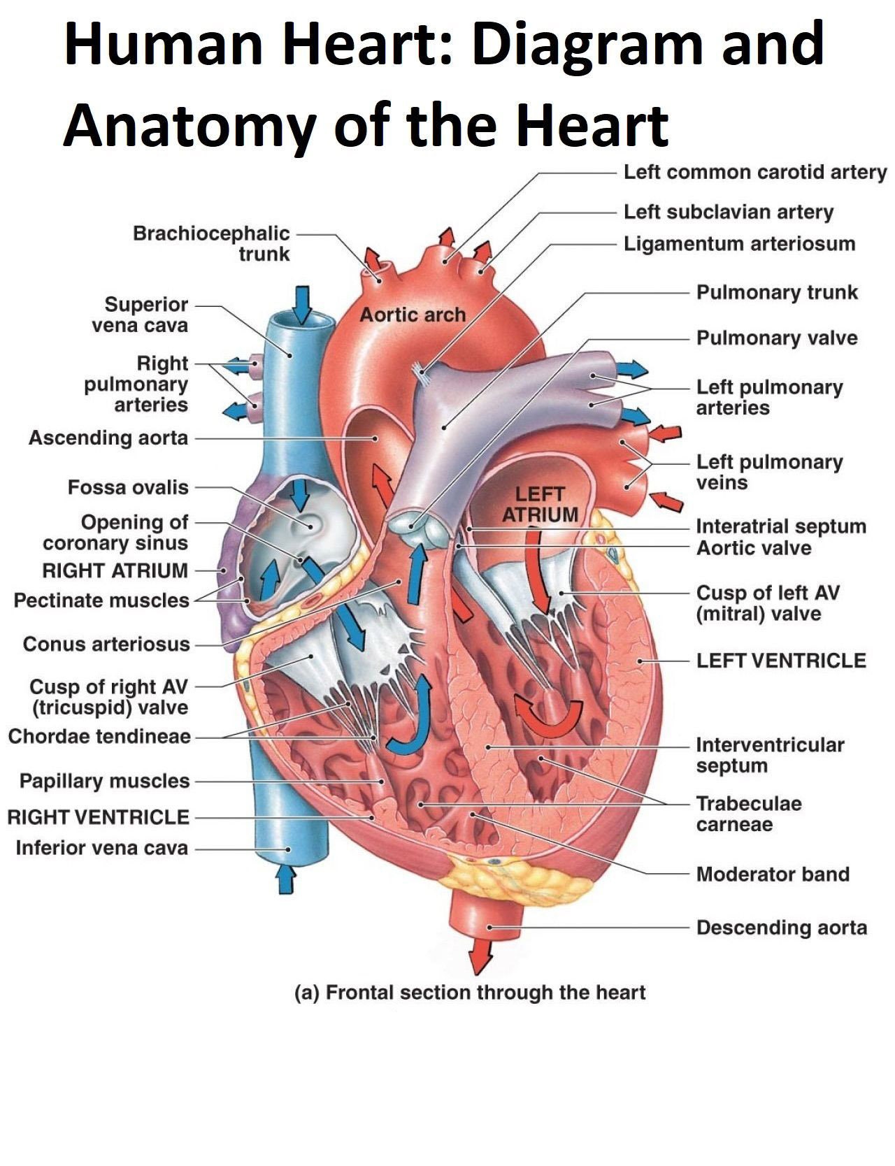
Diagram and Anatomy of the Heart Poster Etsy
This heart was enlarged and displaced the lower lobe of the left lung. the heart is positioned in the chest with 2/3 to the left of midline and the inferior aspect is resting on the diaphragm. The apex of the heart is pointing inferiorly and to the left. The media in this section display the epicardial surface of the heart viewed either from.

External Structure Of Heart Anatomy Diagram
The heart is located in the thoracic cavity medial to the lungs and posterior to the sternum. On its superior end, the base of the heart is attached to the aorta,mycontentbreak pulmonary arteries and veins, and the vena cava. The inferior tip of the heart, known as the apex, rests just superior to the diaphragm.
On Heart Kardiohirurgija.rs
Browse 5,000+ human heart diagram stock photos and images available, or search for heart illustration or pulmonary artery to find more great stock photos and pictures. heart illustration pulmonary artery kidney diagram Sort by: Most popular heart anatomy heart anatomy. Part of the human heart Human Heart Circulatory System

Human heart anatomy. Vector diagram in 2021 Heart anatomy, Human heart anatomy, Human heart
What the Heart Looks Like Español Your heart is in the center of your chest, near your lungs. It has four hollow chambers surrounded by muscle and other heart tissue. The chambers are separated by heart valves, which make sure that the blood keeps flowing in the right direction.

Labeled Diagram Of The Heart And Blood Flow
Heart conditions are among the most common types of disorders affecting people. In the United States, heart disease is the leading cause of death for people of all genders and most ethnic and racial groups. Common conditions that affect your heart include: Atrial fibrillation (Afib): Irregular electrical impulses in your atrium.

Heart anatomy, Heart diagram, Human heart diagram
+ Show all Heart anatomy The heart has five surfaces: base (posterior), diaphragmatic (inferior), sternocostal (anterior), and left and right pulmonary surfaces. It also has several margins: right, left, superior, and inferior: The right margin is the small section of the right atrium that extends between the superior and inferior vena cava .